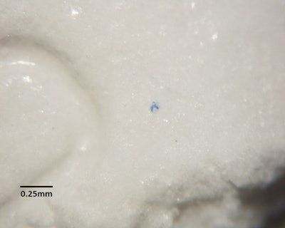
Pharmaceutical packaging is designed to keep the product free from damage and contamination; however, occasionally the packaging itself is the source of product contamination. A pharmaceutical company noticed a problem with several white tablets that displayed small blue defects at random locations on all faces of the tablets. The defects were less than 0.1mm in size, as shown in the photograph here.
When the tablets were cross-sectioned with a razor blade, it was found that the defects were limited to the outer surface of the tablets, and consisted of very thin flakes of a blue substance. The pharmaceutical firm suspected that the defects were fiber fragments from blue cloth wipes or blue hairnets that were used in the process areas of the manufacturing plant. While working in a cleanroom under a stereomicroscope, McCrone removed some of the blue flakes from the tablets by hand by with the aid of fine tungsten needles.
The flakes were pressed onto a salt crystal, and infrared spectra were obtained from the blue flakes and from pieces of cleanroom wipes and hairnets. However, infrared spectra of the blue particles did not match the wipes or hairnets. In addition, the infrared spectra of the blue particles were not of sufficient quality to conclusively identify the blue substance. This sometimes occurs when the particle size is near the detection limit of the infrared system, typically 10-20µm; some of the blue flakes from the tablets were much smaller.
After further discussion with the company, McCrone focused on the desiccant canister that was present in the tablet bottle. The canister was printed with several layers of ink, and the outermost layer in most areas was blue. The ink’s blue color was visually similar to the blue flakes on the tablets. Working under the microscope with a razor and fine needles, McCrone shaved small particles of the blue surface layer of ink from the canister without removing any of the underlying ink layers. The blue ink particles and the blue flakes from the tablets were mounted on a glass slide and analyzed by Raman microspectroscopy. The Raman method is very responsive to pigments and inorganic materials, and can analyze particles as small as 1µm. The Raman spectra showed that the blue ink layer of the ink from the canister ink contained phthalocyanine blue pigment, and the same pigment was the major component of the blue defects on the tablets.
When the pharmaceutical company contacted the supplier of the desiccant canisters, the canister manufacturer was able to correct a formulation problem with the ink, preventing further customer complaints.
Article supplied by Mary Stellmack, senior research chemist, McCrone Associates, Inc., the analytical services division of The McCrone Group. Stellmack co-teaches a course in Infrared Microscopy at Hooke College of Applied Sciences.
